PhotonIcs and Electromagnetics Research Symposium,
also known as Progress In Electromagnetics Research Symposium
PIERS Proceedings
Published: 2015-08-28
Using Diffusion-weighted Images to Identify Brain Tumors
By
Proceedings of 2015 Photonics & Electromagnetics Research Symposium, Prague, July 6 - 9,Page(s)2340-2343
Abstract
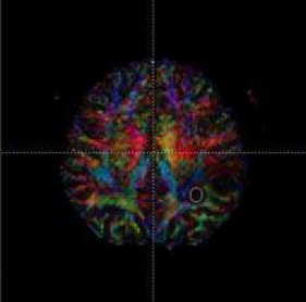
The paper presents an evaluation of the magnetic resonance images of a pathology in the human brain. The experiment involved patients with high-grade glioma tumors in the brain, and an MR tomograph operated by the University Hospital Brno-Bohunice was utilized in the related examinations. In our investigation, we measured images weighted by diffusion. A very interesting technique is diffusion-weighted imaging (DWI), where the measurement sequence comprises a table with 32 vectors of the b-factor orientation. The goal is to perform signal processing in the measured diffusion-weighted images. We proposed a special algorithm for the processing of the DWI image signal. The next step in the procedure was the statistical evaluation of DWI images of healthy and diseased human tissues. We also calculated the brain white matter images, such as those of the fraction anisotropy, RA (relative anisotropy), and VR (volume ratio).
Citation
Karel Bartusek, Petr Marcoň, and Andrea Sprlakova, "Using Diffusion-weighted Images to Identify Brain Tumors," Proceedings of 2015 Photonics & Electromagnetics Research Symposium, Prague, July 6 - 9,Page(s)2340-2343
References