Proceedings of 2015 Photonics & Electromagnetics Research Symposium, Prague, July 6 - 9,Page(s)1861-1864
Abstract
The use of magnetic resonance tomography to scan biological tissues is currently
a very dynamic approach. Based on various image parameters, the method enables us to an-
alyze tissue properties, recognize healthy and pathological tissues, and diagnose the disease or
indicate its progression. These activities are then necessarily accompanied by the processing of
the acquired images. The paper introduces a comparison of statistical tools for the trainable
segmentation of multiparametric data obtained through magnetic resonance tomography. In this
context, the author briefly compares various available tools (Weka, Slicer3D, and RapidMiner)
in view of the input data training and testing, applicability of the classification models, and
ability of the input/output data to be extended with other systems for further processing. The
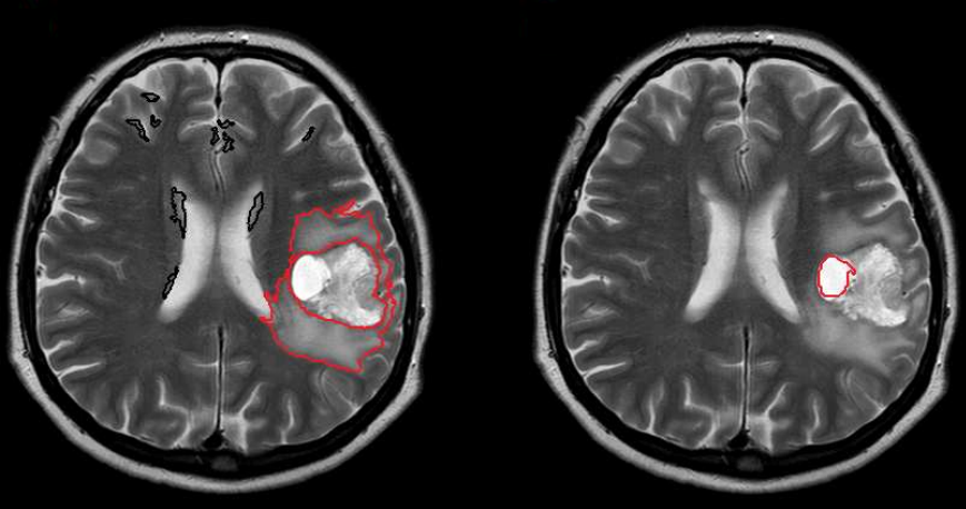
paper also describes as a multiparametric task the segmentation of a brain tumor performed with
real MR data. The source of the data consists in T1 and T2-weighted images. The proposed
segmentation method is carried out within the following phases: data resampling; spatial data
coregistration; definition of the training points; training of the SVM classification model; testing
of the model and interpretation of the classification results.
Citation
Jan Mikulka,
"Multiparametric Biological Tissue Analysis: A Survey of Image Processing Tools," Proceedings of 2015 Photonics & Electromagnetics Research Symposium, Prague, July 6 - 9,Page(s)1861-1864