Proceedings of 2015 Photonics & Electromagnetics Research Symposium, Prague, July 6 - 9,Page(s)1857-1860
Abstract
Using magnetic resonance tomography to scan biological tissues is currently a very
dynamic approach. Based on various image parameters, the method enables us to analyze tissue
properties, recognize healthy and pathological tissues, and diagnose the disease or indicate its
progression. However, the acquired data must be correctly interpreted and visualized by means
of a suitable software tool, such as 3DSlicer (http://www.slicer.org). This well-designed platform
provides an interface between the user and the data available in the popular DICOM format, and
it facilitates very simple 3D visualization of the MR-based data. One of the main advantages of
the open-source software package is undoubtedly its ability to be extended with supplementary
modules, for example the Matlab script. The paper describes the open-source environment with
a focus on Slicer3D and introduces possible extension of this platform with a module for MR data
processing via the three-dimensional, mutiparametric, SVM trainable segmentation method. The
module is freely downloadable. The paper also presents a comparison of the processing results
with respect to the cycle time and the necessary interactivity. Moreover, the author proposes
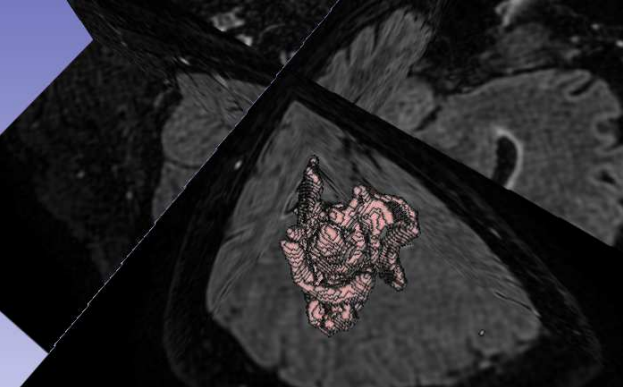
multiparametric segmentation of a brain tumor edema from T1 and T2-weighted images, and the
advantages of the SVM technique are compared with corresponding features of both other fast
segmentation methods and the one-parameter approach.
Citation
Jan Mikulka,
"Matlab Extension for 3DSlicer: A Robust MR Image Processing Tool," Proceedings of 2015 Photonics & Electromagnetics Research Symposium, Prague, July 6 - 9,Page(s)1857-1860