PhotonIcs and Electromagnetics Research Symposium,
also known as Progress In Electromagnetics Research Symposium
PIERS Proceedings
Published: 2015-08-28
Automatic Extraction of Pathological Area in 2D MR Brain Scan
By
Proceedings of 2015 Photonics & Electromagnetics Research Symposium, Prague, July 6 - 9,Page(s)1885-1889
Abstract
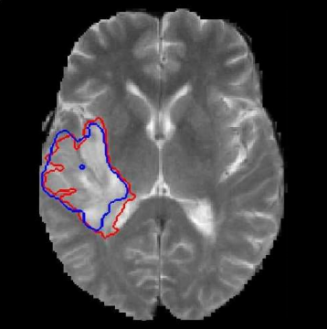
The aim of this work is to propose the fully automated pathological area extraction. The proposed method is based on multi-resolution symmetry analysis and automatic thresholding. The multi-contrast MRI (T2 and FLAIR images) is used. Since the method is based on symmetry, it works for both axial and coronal planes. In these both planes of healthy brain, the approximate left-right symmetry exists and it is used as the prior knowledge for searching the approximate pathology location. The algorithm was tested on 357 axial and 443 coronal real images from publicly available BRATS databases containing 3D brain volumes afflicted by a brain tumor. The results were evaluated by Dice Coefficient (axial: 0.85 ± 0.11, coronal 0.82 ± 0.18) and by Accuracy (axial: 0.96 ± 0.05, coronal 0.94 ± 0.09).
Citation
Karel Bartusek, Pavel Dvorak, and Eva Gescheidtova, "Automatic Extraction of Pathological Area in 2D MR Brain Scan," Proceedings of 2015 Photonics & Electromagnetics Research Symposium, Prague, July 6 - 9,Page(s)1885-1889
References